Meihuayi gives you a high resolution of the principle of TLC
A thin-layer scanner is a special spectrophotometer that can scan spots. It uses visible light or ultraviolet light as a light source, linear scanning or sawtooth scanning of the unfolded spots of the thin-layer plate, and the spot absorbs the monochromatic light of the characteristic wavelength of the component. The remaining monochromatic light is transmitted or reflected or emitted fluorescence, and is integrated by the detector to obtain the content of the substance to be tested in the area of ​​the spot.
principle
Thin-layer chromatography, also known as thin-plate chromatography, is one of the chromatographic methods. It is an important experimental technique for rapid separation and qualitative analysis of small substances. It is a solid-liquid adsorption chromatography. It combines the advantages of column chromatography and paper chromatography. It is suitable for the separation of a small amount of sample (several to a few micrograms or even 0.01 micrograms). On the other hand, when making a thin layer, the adsorption layer is thickened and enlarged. It can also be used to refine samples. This method is especially suitable for substances with low volatility or high temperature changes that cannot be analyzed by gas chromatography. In addition, TLC can be used to track organic reactions and a "pre-test" prior to column chromatography.
Thin layer chromatography (Thin Layer Chromatography) is commonly used in TLC, also known as thin layer chromatography, which belongs to solid-liquid adsorption chromatography. It is a small, fast and simple chromatography method developed in recent years, which combines the advantages of column chromatography and paper chromatography. On the one hand, it is suitable for the separation of small samples (several to several tens of micrograms, or even 0.01 μg); on the other hand, if the adsorption layer is thickened when the thin layer is made, the sample is separated into one line, and the separation can be as many as 500 mg of sample. It can therefore be used to refine the sample. Therefore, the method is particularly suitable for materials that are less volatile or subject to change at higher temperatures and cannot be analyzed by gas chromatography. Further, in the case of performing a chemical reaction, the gradual disappearance of the spot of the raw material is often observed by thin layer chromatography to judge whether or not the reaction is completed.
Thin layer chromatography is to uniformly coat a layer of adsorbent or support agent on the cleaned glass plate (about 10×3cm). After drying and activation, the sample solution is applied to the thin layer plate by capillary tube flattening. The starting line of about 1 cm at one end was cooled or dried, and the thin layer was placed in a developing tank containing a developing agent, and the immersion depth was 0.5 cm. When the leading edge of the developing agent is about 1 cm from the top, the chromatography plate is taken out, dried, sprayed with a color developing agent, or developed under an ultraviolet lamp.
The thin layer chromatograph includes: a thin layer plate sampler, a manual coating station, a dry storage tank, a wash plate holder, and an ultraviolet light detector. It can quickly separate fatty acids, steroids, amino acids, nucleotides, alkaloids and many other substances.
category
1. Centrifugal thin layer chromatography 2. Rod thin layer chromatography 3. Manual thin layer chromatography 4. Dual wavelength thin layer chromatography scanner 5. Automatic thin layer chromatography 6. Rotating thin layer chromatography 7. Full wavelength thin Layer Chromatograph 8. Almighty Thin Layer Chromatograph 9. Diode Array Thin Layer Chromatography Scanner 10. Pressurized Thin Layer Chromatograph 11. Dark Box Thin Layer Chromatography
use
It is suitable for separation, refining, qualitative identification, impurity inspection and content determination of various compounds in the fields of chemistry, chemical engineering, medicine, clinical, agriculture, food, toxicology and so on.
Method of operation
(1) Preparation of thin layer plate Unless otherwise specified, 1 part of stationary phase and 3 parts of water are ground and mixed in one direction in a mortar.
After removing the bubbles on the surface, pour into the applicator and smoothly move the applicator on the glass plate for coating (thickness 0.2~)
0.3mm), remove the thin layer of the glass plate, dry it on the water platform at room temperature, then bake at 110 ° C for 30 minutes, set
Use a desiccant in a dry box for later use. Check the uniformity before use (viewable by transmitted light and reflected light).
(2) Spotting, unless otherwise specified, use a spotter to spot the thin layer, usually a dot, and the baseline is at the bottom.
Side 2.0cm, sample diameter and distance between points are the same as paper chromatography. The distance between spots can be visualized to prevent the detection of the spot.
should. Care must be taken not to damage the surface of the thin layer when spotting.
(3) If the unfolding cylinder is to be saturated with the developing agent in advance, a sufficient amount of developing agent can be added to the cylinder and the wall is
Attach two strips of the same height and width as the cylinder, one end is immersed in the developing agent, and the top of the cylinder is sealed to balance the system or
Operate as specified in the text.
Place the thin plate of the sample into the developing agent of the unwinding cylinder, and immerse the developing agent at a depth of 0.5 from the bottom edge of the thin plate.
~1.0cm (do not immerse the sample in the developing agent), seal the cylinder head, and expand it to the specified distance (usually 10~15cm).
Remove the thin layer plate, dry it, and test according to the regulations under each item.
(4) Scan the chromatographic spots with a thin layer scanner or scan the chromatographic spots directly on a thin layer.
For quantification, thin layer scanning can be used.
The thin layer scanning method can be based on the structural characteristics and usage instructions of various thin layer scanners unless otherwise specified.
In combination with the specific case, the absorption method or the fluorescence method is selected to scan with a dual wavelength or a single wavelength. Due to the impact of thin layer scan results
There are many factors, so the test sample should be tested with the spot of the test sample being linear within a certain concentration range.
The control is scanned on the same thin layer and scanned for comparison and quantification to reduce errors. Various test samples,
Satisfactory results can only be obtained by thin-layer chromatography with good resolution and reproducibility.
Post-elution assay
After the thin layer is unfolded, the adsorbent of the spot is trapped with a doctor blade or a trap, and then eluted with an appropriate organic solvent, and then the content is determined by visible, ultraviolet spectrophotometry, and fluorescence spectrophotometry.
However, the sample spots cannot be sprayed with the color developing agent, so as not to affect the method of determining the position of the spot.
The sample emits fluorescence under ultraviolet light, and the spot can be observed under a UV lamp and the spot position can be removed with a needle.
The color was developed with iodine vapor, and brown spots were treated in the components (the iodine adsorbed and volatilized).
Using the standard control, when the standard point is on the side and the color is measured, the sample is covered to determine the spot position.
fold
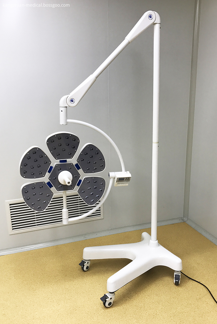
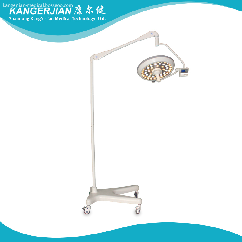
Mobile Type LED Operating Light
Germany imported beads
Imported French lens
mould Die-casting Eight edge type Revolving arm
Optional emergency power supply≥3 hours


Product features:
1.Ideal cold light effects.
Using the new LED cold light source, energy saving and environmental protection and long service life up to 80,000hours or more. Temperature increase over surgeon`s head < 1℃.
LED do not engender infrared ray and ultraviolet radiation, it doesn`t have the temperature rise and tissue damage caused by halogen shadowless light, can accelerate the wound healing after surgery, and has no Radiation pollution.
LED color temperature constant, soft, very close to the natural sun light.
2.Excellent shadowless effect
Lamp with the most scientific radian,Multi point light source design, so that more fullness of the light spot,When the lamps are partially occluded, also can achieve perfect shadowless effect.
Lamp panel radius of gyration: ≥182cm, the lamp can be pulled to vertical floor, convenient to any angle illumination.
3.Excellent deep lighting
4.Advanced control system
The use of liquid crystal display button control, to meet the needs of the medical staff of different patients with the brightness of the operation.
It offers illuminance memory function.
5.Universal suspension system
Rotating arm, a new type of alloy material is made of eight edge type.
Balanced system using imported arm module, more than 5 group universal joints, every cantilever must has more than 3 joints which can be rotated in 360°, The structure is light, easy to manipulate, accurate positioning, can provide the maximum range of regulation.
The equipped with fatigue correcting unit and fix position hand handle device, easy to fix position after long time use.
6.Modern laminar lamp
The thickest part of lamp-chimney is not more than 10cm.
The lamp-chimney is made of ABS, The handle on the central of lamp can be detachable, can take high temperature (≤ 134°C) sterilization treatment, easily adjust, flexible fixed.
Mobile Type LED Operating Light,Mobile Operating Light,Mobile LED Surgical Light,Movable LED Operating Lamp
Shandong qufu healthyou Medical Technology co.,Ltd , https://www.kangerjianmedical.com