Application examples of myoelectric surface electrodes
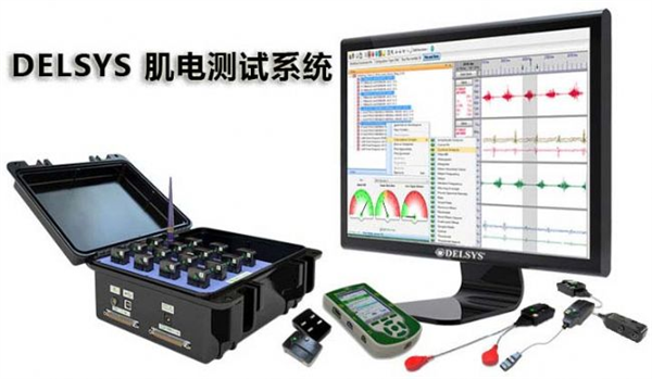
The anterior arm muscle FDS sEMG signal was recorded by array electrode, the RMS value of sEMG signal was extracted, and the correlation with the index finger strength level was analyzed to study the recruitment of motor units at different anatomical locations.
1 method
There were 8 college student volunteers, including 4 males and 4 females. The subjects were between 20 and 24 years old. They were healthy and did not have high-intensity exercise two days before the experiment. There was no motor neurological disease. The process is known.
Experimental content The subject's palm was down, the thumb was adducted, and the sensor was placed on the sensor from 1 to 4, and 1012 different levels of single-finger force tracking experiments were performed as required. During the experiment, the target is provided with the target power curve, and the actual finger force is feedback in real time, so that it tries to imitate the target line to complete the task, complete a round of 6 1012 N force tracking experiment as a group, repeat 5 minutes before the experiment, Each subject has a training session familiar with the experimental process to avoid the adaptability of the subjects in the experiment. The order in which the test is completed is random.
The experimental equipment and parameters were recorded by surface array electrode and RM6280C multi-channel physiological parameter recorder. The surface electromyography signal of the forearm finger shallow flexor (FDS) and the output voltage of the force sensor were used. The electrode was the center distance of each electrode composed of a 2 mm diameter gold-plated round electrode. It is 3mm, and the sampling rate of the signal attached to the front recorder in the direction of the muscle fiber is set to 2000 Hz.
2 data processing
The sEMG signal was analyzed in the time domain using the experiment. The signal segment is selected according to the calibration result in the recording software of the physiological recorder 6280, and the output voltage of the finger force sensor is converted into a power curve, and the power plateau with a length of 1 25 N% is selected, and the segment sEMG signal is used for analysis.
Filtering In Matlab7.0, the original sEMG number is bandpass filtered with an elliptical filter to calculate the RMS value of the sEMG signal after filtering. First, 500 points is used as a time window, and the windows do not overlap. The filtered sEMG signal is divided into 4 segments, and the RMS value of each segment is calculated separately.
3 results
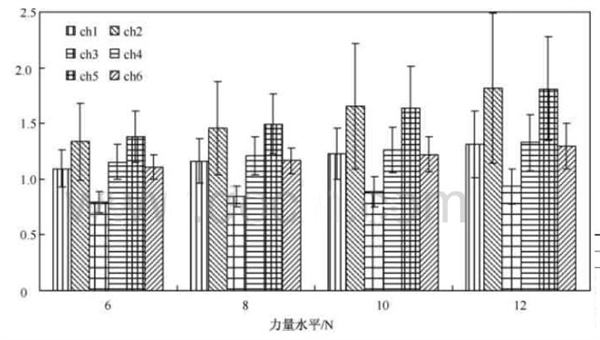
The RMS of all actions for each subject was calculated and then the eigenvalue data repeated 5 times for each force level was averaged. The graph shows the amplitude variation of the RMS value of the sEMG signal for each channel at different power levels.
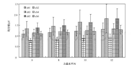
RMS value of each channel sEMG signal varies with power level
Figure 2 shows the variation of the RMS level of the sEMG signal recorded at different electrode point locations. It can be seen that in the index finger activity mode, the sEMG signal recorded by channel 2 is most sensitive to the change in force, the channel is 5 times, and the remaining four channels are RMS. Less sensitive to power. The RMS of Channel 2 and Channel 5 is almost twice as sensitive as the other channels.
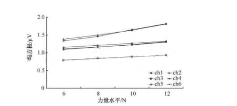
RMS of each channel sEMG signal with force level curve
4 discussion
The RMS of the sEMG signal in each channel shows an increasing trend with increasing power level. It is found by observation that the RMS of the sEMG signal recorded at different electrode points increases with the increase of the power level.
Reiners et al. observed that the firing rate of muscle motor units increased with the increase of muscle strength. When the muscle contraction force was small, the low threshold motor units were recruited, and the firing rate was lower, resulting in higher action potentials. Small; when the muscle contraction strength is large, the higher threshold motor unit is raised faster than the raised threshold exercise unit, and is also irregular, and the measured action potential of the electrode is larger [12].
In addition, the amplitude (RMS) of the sEMG signal increases as the force increases with the conventional two-electrode structure.
This indicates that the electrical activity of high-threshold, high-distribution exercise units recruited with increasing levels of strength through the conduction and synthesis of muscle tissue and skin is also significantly manifested as an increase in the amplitude of the sEMG signal; this study was performed by array electrodes. The two-dimensional sEMG signal further confirms that the motor unit recruitment mode and its electrical activity level related to the power level are also reflected in the changes in the electrical activity intensity at different anatomical locations on the muscle surface, that is, the sEMG with different anatomical positions on the muscle surface as the muscle contraction strength increases. The amplitude will be enhanced synchronously.
The nerve fiber produces excitement and conduction in accordance with the law of “0†or “1â€. There is only “yes†or “noâ€. There is no strong or weak point. Why do you feel strong and weak when you are stimulating? Forbearance? Because of the negative pulse, it actually stimulates multiple bundles of nerve fibers to produce excitation and conduction together, such as fibers of different thickness, incoming and outgoing fibers, and also stimulate muscle contraction, which in turn stimulates other receptors ( Including pain receptors) signals the center, so the feeling is complicated.
2 There are differences in RMS between different spatial anatomical structures of FDS
It can be observed from Fig. 2 that under the same force level, the RMS difference of the sEMG signal recorded by the electrodes at different spatial anatomical structures of the FDS is large.
Figure 3 shows the schematic diagram of the placement of the array electrodes on the superficial flexors in the experiment. Table 1 shows the mean and variance of the RMS of the sEMG signals in each channel. When the muscles contract with a certain force, the corresponding motor units are recruited, and the action potentials are generated by the nerves. The dominating region is transported along the muscle fibers to both sides, the action potential near the innervation region and the tendon is lower, and the action potential in the intermediate region is relatively larger.

It can be seen from Fig. 1 that the RMS of all channel sEMG signals increases with the increase of the force level regardless of the influence of different FDS anatomical positions. At the same time, the RMS amplitude of the sEMG signal recorded by the electrodes in different spatial anatomical locations of the FDS is different. The RMS amplitudes of the sEMG signals recorded by each channel vary greatly. Table 1 shows the mean and standard deviation of the RMS values ​​of the sEMG signals for each channel. The mean and standard deviation and variance of the RMS amplitudes of the channels 2 and 5 can be observed. Both are larger than other channels.
5 Conclusion
In this study, the array electrode was used to collect FDS high-density sEMG signals from the index finger single-finger force output experiment, and the sEMG signal RMS was extracted to analyze the change with the force level.
The results show that the RMS amplitude shows an increasing trend with the increase of finger strength level, which can be used as the characteristic value of sEMG signal to reflect the level of muscle activity;
FDS not only has different functional partitions, but for the same functional partition, different anatomical locations participate in different degrees of finger activity control.
Although this study is a small sample size exploration study, it is confirmed that the array electrode can be used to detect the spatial information of FDS myoelectric activity, estimate the spatial activation characteristics of FDS and the control mode of the finger, in order to further study the spatial activity pattern of the forearm muscle. Technical Support.

Coffee Table Parts,Office Wood Desk Customized,Plastic Wood Coffee Table Customized,Wooden Low Coffee Table
SHENZHEN CHANGJIANG FURNITURE CO., LTD , https://www.findcjf.com